|
This time the influence of tetravalent dopants such as Si4+, Sn4+, Ti4+, and Zr4+ on hematite nanostructure for enhanced photoelectrochemical water splitting is reported. The photoactivity of the doped photoanodes at 1.23 V RHE follows the order Zr > Sn > Ti > Si. The work was performed in collaboration with Prof. Jum Suk Jang (Chonbuk National University, Korea), and the results are published in the journal of Applied Surface Science. A. Subramanian, E. Gracia-Espino, A. Annamalai, H. H. Lee, S Y. Lee, S. H. Choi, and J. S. Jang. Applied Surface Science (2017). DOI: 10.1016/j.apsusc.2017.09.042 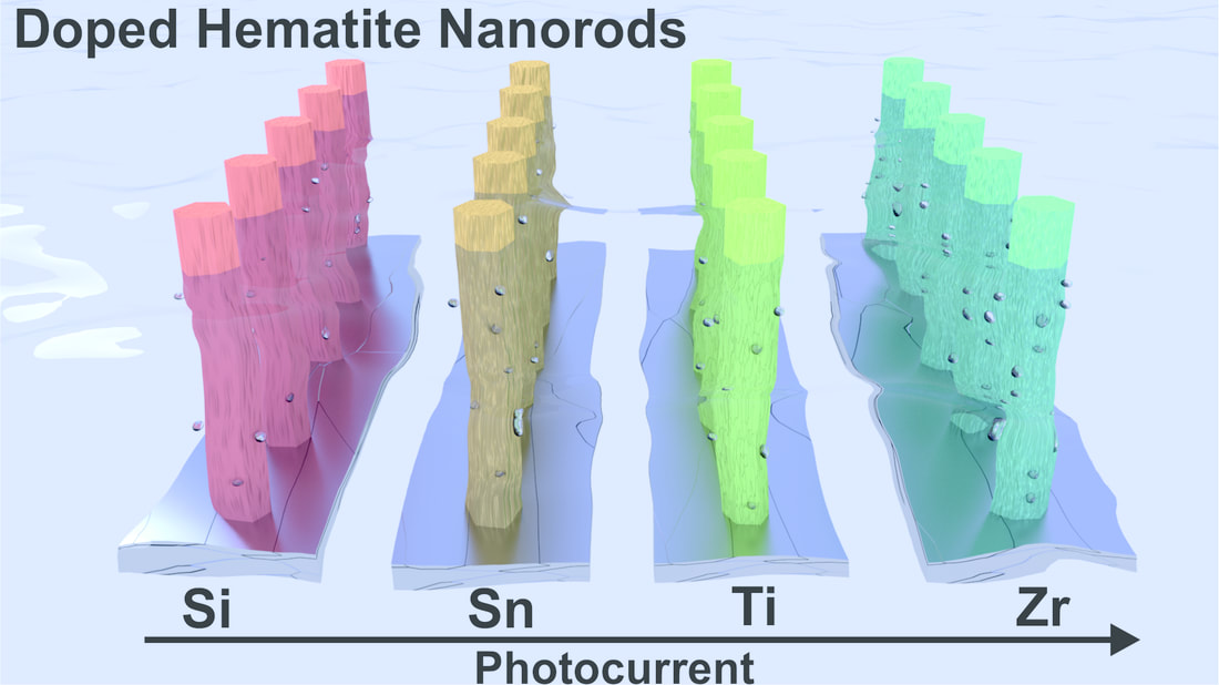 Abstract In this paper, the influence of tetravalent dopants such as Si4+, Sn4+, Ti4+, and Zr4+ on the hematite (α-Fe2O3) nanostructure for enhanced photoelectrochemical (PEC) water splitting are reported. The tetravalent doping was performed on hydrothermally grown akaganeite (β-FeOOH) nanorods on FTO (fluorine-doped tin-oxide) substrates via a simple dipping method for which the respective metal-precursor solution was used, followed by a high-temperature (800° C) sintering in a box furnace. The photocurrent density for the pristine (hematite) photoanode is ∼0.81 mA/cm2 at 1.23 VRHE, with an onset potential of 0.72 VRHE; however, the tetravalent dopants on the hematite nanostructures alter the properties of the pristine photoanode. The Si4+-doped hematite photoanode showed a slight photocurrent increment without a changing of the onset potential of the pristine photoanode. The Sn4+- and Ti4+-doped hematite photoanodes, however, showed an anodic shift of the onset potential with the photocurrent increment at a higher applied potential. Interestingly, the Zr4+-doped hematite photoanode exhibited an onset potential that is similar to those of the pristine and Si4+-doped hematite, but a larger photocurrent density that is similar to those of the Sn4+- and Ti4+-doped photoanodes was recorded. The photoactivity of the doped photoanodes at 1.23 VRHE follows the order Zr > Sn > Ti > Si. The onset-potential shifts of the doped photoanodes were investigated using the Ab initio calculations that are well correlated with the experimental data. X-ray diffraction (XRD) and scanning-electron microscopy (FESEM) revealed that both the crystalline phase of the hematite and the nanorod morphology were preserved after the doping procedure. X-ray photoelectron spectroscopy (XPS) confirmed the presence of the tetravalent dopants on the hematite nanostructure. The charge-transfer resistance at the various interfaces of the doped photoanodes was studied using impedance spectroscopy. The doping on the hematite photoanodes was confirmed using the Mott-Schottky (MS) analysis.
0 Comments
Your comment will be posted after it is approved.
Leave a Reply. |
Nano for Energy group
Categories
All
Featured publications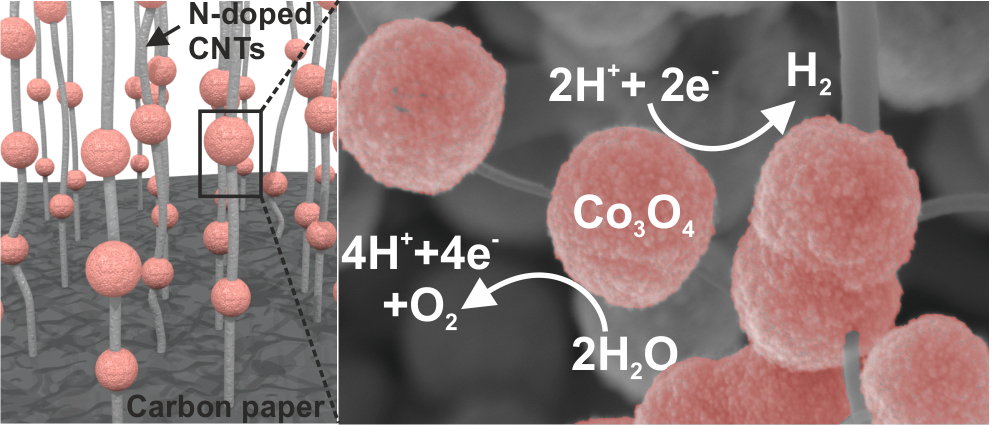
Comprehensive Study of an Earth-Abundant Bifunctional 3D Electrode for Efficient Water Electrolysis in Alkaline Medium.
ACS Appl. Mater. Interfaces, 2015, 7, 28148 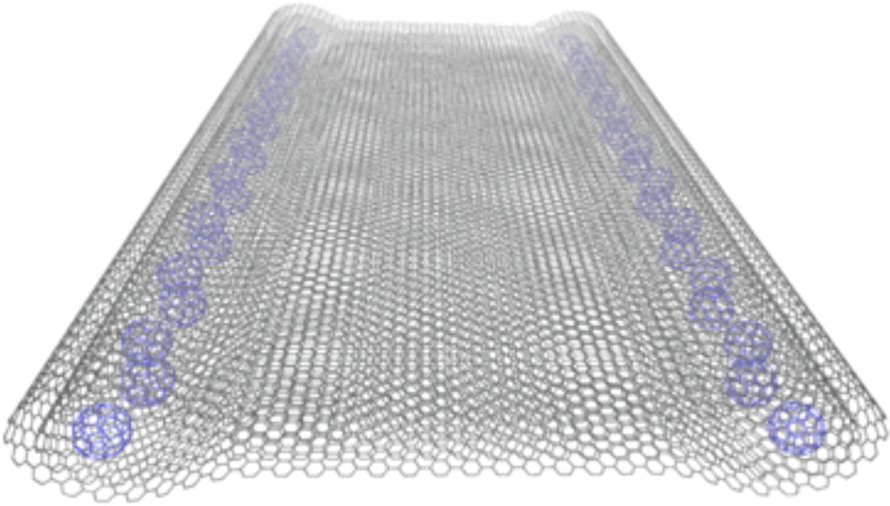
Fabrication of One-Dimensional Zigzag [6,6]-Phenyl-C61-Butyric Acid Methyl Ester Nanoribbons from Two-Dimensional Nanosheets.
ACS Nano, 2015, 9, 10516 
Hierarchical self-assembled structures based on nitrogen-doped carbon nanotubes as advanced negative electrodes for Li-ion batteries and 3D microbatteries.
J. P. Sources, 2015, 279, 581 
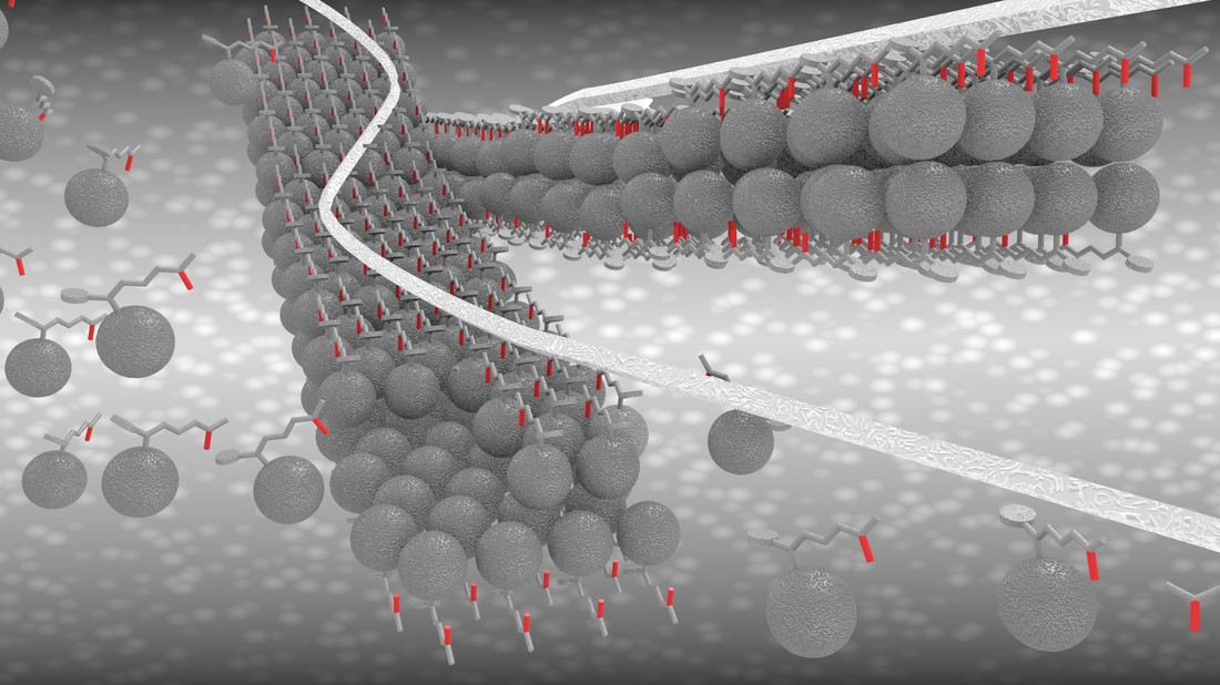
.Self-Assembly Synthesis of Decorated Nitrogen-Doped Carbon Nanotubes with ZnO Nanoparticles: Anchoring Mechanism and the Effects of Sulfur.
J. Phys. Chem. C, 120, 27849 (2016) 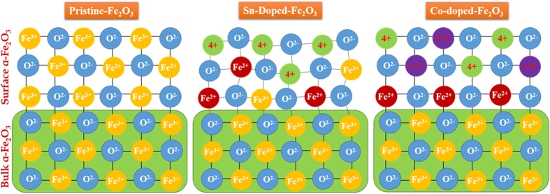
Sn/Be Sequentially co-doped Hematite Photoanodes for Enhanced Photoelectrochemical Water Oxidation: Effect of Be2+ as co-dopant.
Sci Rep. 2016; 6: 23183. 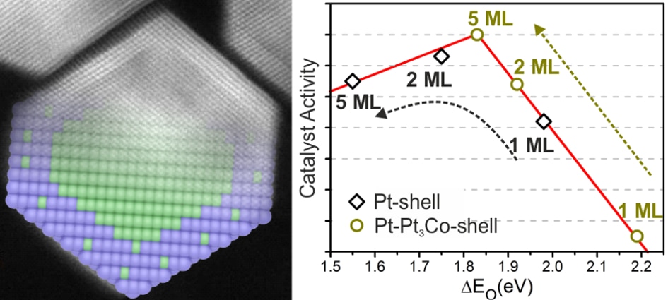
Atomistic understanding of the origin of high oxygen reduction electrocatalytic activity of cuboctahedral Pt3Co–Pt core–shell nanoparticles.
Catal. Sci. Technol., 2016, 6, 1393-1401 
Photocatalytic reduction of CO2 with H2O over modified TiO2 nanofibers: Understanding the reduction pathway.
Nano Res. (2016) 9: 1956. |
 RSS Feed